
In 2024-2025, I collaborated with Heran Getachew1 and Marcela Garita-Hernandez1 to design scientific figures to illustrate their publication Getachew, H et al. (2025) Advances and optimization strategies in prime editing of human pluripotent stem cells (https://doi.org/10.1016/j.tibtech.2025.06.017).
In terms of tools, I was using Inkscape to create a single large SVG file containing a separate canvas per figure that I would rasterize as high-resolution TIFF (all at once or individually).
Just below, you’ll find some of the final figures I made.
1 – Ocular Genomics Institute, Massachusetts Eye and Ear Infirmary, Department of Ophthalmology, Harvard Medical School, Boston, MA 02114, USA
Fig.1 – Key components of prime editing:
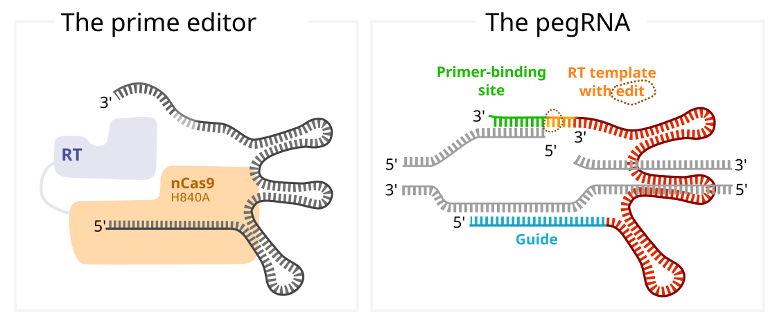
Abbreviation: RT, reverse transcriptase. Figure produced in collaboration with 42Borgata. (Published in Trends in Biotechnology)
Fig.2 – Mechanism of prime editing:
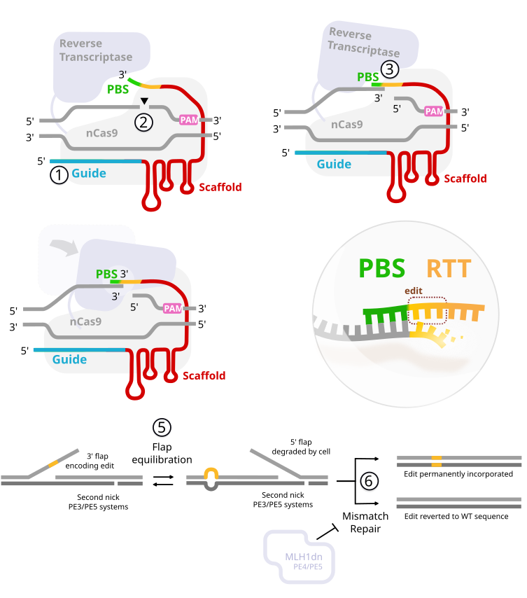
Abbreviations: PAM, protospacer adjacent motif; PBS, primer-binding site; RTT, reverse transcription template; WT, wild type. Figure produced in collaboration with 42Borgata. (Published in Trends in Biotechnology)
Fig.3 – Prime editors
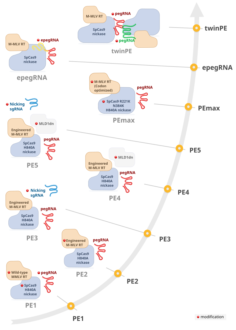
Abbreviations: prime-editing guide RNA (pegRNA); RT, reverse transcriptase; sgRNA, single-guide RNA. Figure produced in collaboration with 42Borgata. (Published in Trends in Biotechnology)